
Eye Disease Management
Comprehensive Disease Management Solutions for Optimal Eye Health
The most important part of your eye exam is the ocular health evaluation. At Stoughton Eye Care & Eyewear, we believe prevention is the best medicine.
We use cutting-edge technology that goes beyond your basic eye exam to help detect eye disease such as diabetic retinopathy, age-related macular degeneration, glaucoma, and cataracts at their earliest stages. Our goal is to use the best resources available to prevent vision loss and maintain your eye health.

Advanced Microscopy
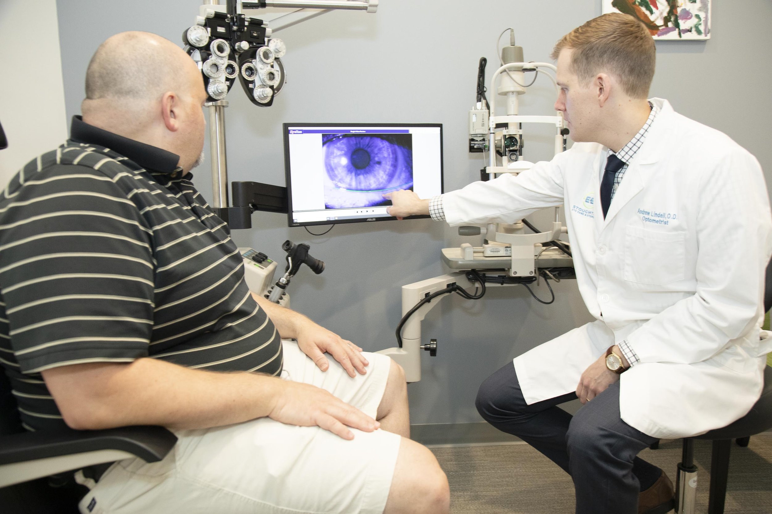
To examine your eyes we will use our elaborate microscopes that allow for digital imaging and videos to be taken of your eye. This allows us to properly document changes such as cataracts, dry eye, corneal disorders and monitor your treatment progress. We will be able to show you exactly what is going on with your eye health so you have a clear understanding.
OCT of Macular Degeneration
OCT angiography of optic nerve for glaucoma testing
Optical Coherence Tomography (OCT)
Avanti OCT and OCT angiography are non-invasive imaging tests used to view the layers of the retina, evaluate the optic nerve health and measure the blood flow of the eye. This allow us to detect diseases such as:
Dry and Wet Age Related Macular Degeneration, (AMD)
Diabetic Macular Edema
Glaucoma
Diabetic Retinopathy
Hypertension Retinopathy
Macular Puckering
Macular Holes
Macular edema
OCT is also utilized to monitor disease progression quickly and accurately.
Visual field of hemianopsia from brain tumor
Visual field showing vision loss from advanced glaucoma
Visual Field Evaluation
We perform a quick, easy and comfortable evaluation of how far you can see in your peripheral and central vision. This helps to evaluate you for blind or missing spots in your vision.
The location and size of the missing spots can be caused by different eye diseases and brain disorders. Systemic diseases such as multiple sclerosis, thyroid eye disease or Grave’s disease, pituitary gland disorders, stroke, brain tumors and certain high risk medication such as Plaquenil can all cause missing spots in your vision.
Optomap image showing changes in age related macular degeneration
2 eyes with diabetic retinopathy captured with Optomap
Optomap
Optomap is an advanced fundus imaging technology that allows us to evaluate the retina, or the back of the eye, out to 200 degrees. The images are easy and quick to take for patients of all ages.
With a traditional dilation, quick limited views of the retina are obtained and can be hindered by patients' ability to keep their eyes open for evaluation with our bright lights. The digital ultra-wide field image allows detailed evaluation of the fine blood vessels, optic nerve, macula and the peripheral retina and the ability to utilize diagnostic measurements and provide proper documentation to evaluate for disease progression over time.
Advanced retina evaluation can help to find the first signs of diseases including stroke, diabetes, high blood pressure, and some cancers, even before you have symptoms. It can help to find early retina thinning, tears, holes and breaks that can be treated to prevent the onset of retinal detachment.